mbilical Hernia: Understanding, Diagnosis, and Treatment
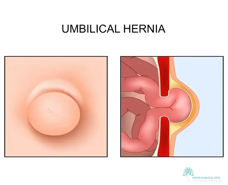
Umbilical hernias occur when tissue—often part of the intestine or fatty tissue—protrudes through a weakness or opening in the abdominal muscles near the belly button. Although commonly encountered in both children and adults, many people remain unaware of their potential to cause discomfort or lead to complications if left untreated. Learning about umbilical hernias—their causes, warning signs, diagnostic process, and treatment options—is key to managing this condition effectively.
Causes of Umbilical Hernias
Umbilical hernias can result from either congenital or acquired factors, with each group displaying distinct characteristics.
Congenital Factors
Developmental Weakness: Some infants are born with weak abdominal muscles, making it easier for internal tissues to push through the natural opening where the umbilical cord once passed.
Delayed Closure: Typically, the opening in the abdominal wall should close shortly after birth. However, if it remains open, an umbilical hernia can develop.
Higher Risk in Preterm or Low-Birth-Weight Infants: Premature infants and those with low birth weights are more likely to experience this condition.
Acquired Factors
In adults, umbilical hernias are often acquired due to increased pressure inside the abdomen:
Obesity: Excess body weight creates additional pressure on the abdominal wall, making it easier for a hernia to form.
Pregnancy: Multiple or prolonged pregnancies stretch and weaken the abdominal muscles.
Heavy Lifting and Straining: Repeated or intense lifting and straining can gradually cause a weak spot near the belly button.
Previous Abdominal Surgery: Incisions or scars from past surgeries can become areas of weakness in the muscle wall.
Understanding these causes helps in recognizing risk factors and adopting preventive measures.
Recognizing the Symptoms
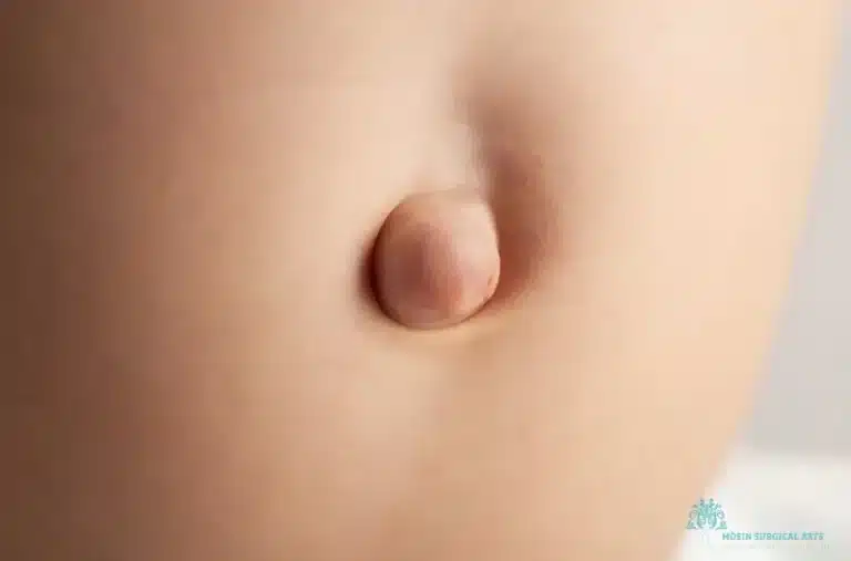
Noticeable Bulge Near the Belly Button
The hallmark of an umbilical hernia is a visible or palpable bulge at or near the navel. This bulge:
Can become more prominent during activities that increase abdominal pressure, such as coughing, crying, or straining.
May reduce or disappear when lying down or resting.
Discomfort and Pain
While many umbilical hernias are painless, discomfort or pain may develop if:
The hernia becomes incarcerated (trapped) within the abdominal wall.
There is pressure on nearby nerves.
The size of the hernia increases over time.
Changes During Activity
The size and prominence of the bulge may fluctuate with physical activity. For instance:
During Strain: The bulge can enlarge, causing temporary discomfort.
At Rest: The hernia may appear smaller and less concerning.
Diagnosis: A Step-by-Step Approach
Accurate diagnosis of an umbilical hernia is vital to determine the proper treatment. The process typically involves:
Physical Examination
Inspection: Your healthcare provider will look for a protruding lump near the belly button.
Palpation: They may ask you to cough or stand to observe any changes in the size of the bulge.
Assessment: The doctor evaluates the hernia for tenderness, size, and reducibility (whether the protruding tissue can be pushed back into the abdominal cavity).
Imaging Studies
If the physical exam is inconclusive or if complications are suspected, additional tests may include:
Ultrasound: A non-invasive method using sound waves to visualize the hernia.
CT Scan: Offers detailed images of the abdominal wall and can assess the size and contents of the hernia.
MRI: Occasionally used to provide a clearer picture if other imaging tests are insufficient.
Biopsy (if necessary)
In rare cases, tissue samples may be taken to rule out more serious conditions, such as malignancies.
Treatment Options for Umbilical Hernias
Treatment strategies depend on the hernia’s severity, the symptoms it produces, and the patient’s overall health.
Surgical Repair
Surgical intervention is often recommended for larger or symptomatic hernias. Two primary surgical techniques include:
Open Surgery
Procedure: A single incision is made near the hernia, the protruding tissue is gently pushed back, and the muscle defect is closed. Often, a mesh is placed to reinforce the area.
Benefits: Effective for larger hernias and provides a durable repair.
Considerations: Involves a longer recovery period and a larger incision.
Laparoscopic Surgery
Procedure: Small incisions are made to insert a camera and surgical instruments. The repair is performed minimally invasively, often including mesh placement.
Benefits: Quicker recovery time and smaller scars.
Considerations: Requires specialized equipment and expertise.
Non-Surgical Management
For small, asymptomatic hernias, non-surgical management may be an option:
Observation: Regular check-ups to monitor the hernia.
Lifestyle Modifications: Avoiding heavy lifting and managing weight to reduce abdominal pressure.
Supportive Devices: Wearing a supportive belt may temporarily alleviate discomfort, though it does not correct the defect.
Treatment Comparison Table
Surgical Technique
Benefits
Considerations
Open Surgery
Durable repair; effective for larger defects
Larger incision; longer recovery
Laparoscopic Surgery
Minimally invasive; quicker recovery
Requires specialized training; may not be ideal for very large hernias
Complications of Untreated Umbilical Hernias
Ignoring an umbilical hernia can lead to serious complications:
Incarceration: The hernia becomes trapped and cannot be reduced back into the abdomen, causing pain and swelling.
Strangulation: The blood supply to the herniated tissue is cut off, leading to tissue death (gangrene) and requiring emergency surgery.
Complications Table
Complication
Symptoms
Risk Level
Incarceration
Increased pain, swelling, vomiting
High
Strangulation
Severe pain, fever, discoloration
Very High (emergency)
Early diagnosis and treatment are essential to avoid these life-threatening conditions.
Recovery and Post-Operative Care
After Surgical Repair
Recovery time varies but typically includes:
Immediate Recovery: A short hospital stay (or outpatient recovery) with initial pain management.
Activity Restrictions: Limited physical activity for 4–6 weeks, with gradual return to normal activity as advised.
Wound Care: Careful cleaning and dressing of the incision site to prevent infection.
Follow-Up: Regular postoperative visits to ensure proper healing and monitor for recurrence.
Home Care Guidelines
Rest and Mobility: Engage in light walking to support circulation, but avoid heavy lifting.
Hygiene and Support: Maintain the cleanliness of the area and use supportive garments as directed.
Monitor for Changes: Keep an eye out for any signs of infection or recurrence, such as increased pain or a new bulge.
Preventing Recurrence
Preventive strategies can help reduce the risk of the hernia recurring after surgical repair:
Lifestyle Modifications
Modification
Benefit
Maintain a Healthy Weight
Reduces abdominal pressure
Avoid Heavy Lifting
Decreases strain on the muscle wall
Core Strengthening Exercises
Strengthens the abdominal muscles
Balanced Diet
Prevents constipation and undue strain
Regular Monitoring
Follow-Up Care: Consistent check-ups with your healthcare provider can catch early signs of recurrence.
Symptom Awareness: Stay alert for any changes in the abdominal area and report concerns promptly.
Final Thoughts: Expert Umbilical Hernia Repair
Umbilical hernias are common yet potentially serious conditions if complications occur. Whether congenital in infants or acquired in adults, understanding the risks and treatment options is essential. At Dr. Babak Moein’s practice, specialized care combines expert surgical techniques with comprehensive follow-up to ensure the best possible outcomes. If you notice any signs of an umbilical hernia or have concerns about your health, early consultation is the key to effective management.
For personalized advice and expert treatment in Los Angeles, CA, contact Dr. Babak Moein today at (310) 455-8020. Take the first step toward restoring your health and confidence with professional umbilical hernia repair.
This updated article provides a new perspective on the causes, symptoms, diagnosis, and management of umbilical hernias, ensuring that prospective patients receive detailed and current information about their condition.